 Brain 2 teaches the form, function, and response mechanisms of the brain's various components and offers hands-on techniques to effectively release many primary restrictions that can affect the whole body. In this class, we will learn different ways to find a primary lesion in the brain, and we will focus a little more on the spinal cord's grey and white matter.
Class Length: 3 days
Conflict of Interest: All classes presented by Chikly Health Institute have no financial conflict of interest.
CHI is not sponsored by outside organizations or corporations.
Please read "Our Policies" for more information: https://chiklyinstitute.com/Policies
• Contact Continuing Education (CE) Hours Total: 18 CEUs for massage therapists - NCBTMB Approved Provider # 451238-10
NCBTMB CEUs are accepted in every US state for NCBTMB certification renewal.
Most states accept NCBTMB for license renewal but not all. We are also approved for NY state.
Please look here for more information: http://www.ncbtmb.org/map/requirements-map.
Because certification and license renewal policies vary from state to state, it's important for you to make sure the CEUs are accepted wherever you practice. Therefore, please be aware that this information may not apply in your state.
Check your state’s website at: http://www.ncbtmb.org/regulators/state-info.
Alberta massage therapists: Members of the RMTA will receive 15 Continuing Education Credits (CEC) upon the submission of a certificate of completion for each course.
• 18 hours approved by the Massage Therapy Association of Manitoba, Canada (MTAM)
• 18 hours approved by the Certified Registered Massage Therapy Association of Alberta, Canada (CRMTA)
We are in the process of providing Continuing education for numerous other professions. Please check back to this page later as we will post all updates.
Schedule (subject to change)
Day One:
8:30 Registration
9:00 - 11:00 Introduction, teachers, students, teaching assistants, and facilitator. Teaching material
Review of some Brain level 1 techniques
Reticular formation (RF) / Reticular Alarm System (RAS): the median, medial and lateral column of the RF.
11:00 - 11:15 Break
11:15 - 12:30 Ventricles of the brain: fluid assessment and treatment
Corpus Callosum: assessment and treatment
12:30 - 2:00 Lunch
2:00 - 3:30 Anterior Commissure: assessment and treatment
The commissure of the Fornix
3:30 - 3:45 Break / group discussion
3:45 - 5:30 Basal Nuclei / Internal Capsule: motor/coordination/balance assessment. Applications in motor deficit, fine motor skills.
Day Two:
9:00 - 11:00 Questions and answers
Thalamus afferents, applications to physical body lesions
11:00 - 11:15 Break / group discussion
11:15 - 12:30 Release of the superior, middle, and inferior peduncles of the cerebellum: fascia and fluid approach.
12:30 - 2:00 Lunch
2:00 - 3:00 Tissue trauma and hands-on downregulation of the RAS and clinical cases
3:00 - 3:30 RAS Clinical cases
3:30 - 3:45 Break / group discussion
3:45 - 5:30 Finding dominant lesions in the CNS: fascia and fluid approaches.
Day Three:
9:00 - 10:30 Questions and answers
Release of Spinal Cord tensions: fascia and fluid approach.
10:30 - 10:45 Break / group discussion
10: 45 - 12:45 Caudal meningeal attachments of the spinal cord: Filum terminale internum and externum
12:45 - 2:00 Lunch
2:00 - 3:30 Cephalic meningeal attachments of the spinal cord: occipital and cervical dural attachments. Foramen Magnum fascial release
Review.
Take home Protocol
Final questions and answers
Self-Reflection and identification of changes for practitioner’s practice
LEARNER OBJECTIVES:
- By the end of the course participants will be able to correctly demonstrate on a live person how to alleviate physical symptoms of Reticular Alarm System hyperactivity
- By the end of the course participants will be able to correctly assess on a live person fluid dysfunctions of the Ventricles
- By the end of the course participants will be able to correctly demonstrate on a live person how to release dysfunctions of the Corpus Callosum
- By the end of the course participants will be able to correctly demonstrate on a live person how to release dysfunctions of the Anterior Commissure
- By the end of the course participants will be able to correctly assess on a live person fluid dysfunctions of the Internal Capsule
- By the end of the course participants will be able to correctly assess on a live person dysfunctions of the Thalamus
- By the end of the course participants will be able to correctly demonstrate on a live person how to release the peduncles of the Cerebellum
- Based on a fascia and fluid approach participants will be able by the end of the course to correctly design on a live person a proper treatment plan in relation to CNS dysfunctions
- By the end of the course participants will be able to correctly demonstrate on a live person how to release mechanical dysfunctions of the spinal cord
- By the end of the course participants will be able to correctly demonstrate on a live person how to release caudal meningeal attachments of the spinal cord
- By the end of the course participants will be able to correctly demonstrate on a live person how to release cephalic meningeal attachments of the spinal cord.
INSTRUCTIONAL METHODS
- Lecture
- Study Guide
- Question & Answer
- PowerPoint Slides
- Demonstration
- Practice sessions
- Review
B1: Brain Tissue, Nuclei, Fluid & Autonomic Nervous System, plus at least three (3) months of practice after attending B1.
An in-depth advanced study of anatomical terms and concepts is necessary; a study list is provided below.
Anatomical/Physiological Terms
1. The DVD "Dissection of the Brain" and Spinal Cord is a great preparation for the Brain 2 class.
2. Be sure you understand the following words and, as applicable, know precisely where these structures are located in the body.
For this class, it is more important to know their 3-dimensional location and relationship with one another, than their classically described physiology.
- Reticular alarm system: median (raphe nuclei), medial and lateral columns, locus ceruleus, sulcus limitans, rhomboid fossa.
- Frontal and occipital forceps of the corpus callosum
- Internal capsule
- Anterior commissure, medial and lateral olfactory striae, posterior commissure
- The commissure of the fornix (psalterium or lyre of David)
- Mamillothalamic tract
- Posterior limb of the internal capsule, pyramidal decussation, corticospinal tracts
- Dentate gyrus, gyrus fasciolaris
- All nuclei of the thalamus, including midline, medial dorsal (MD), all lateral and all ventral, intralaminar nuclei (centromedian, central lateral, parafascicular), etc.
- Nucleus accumbens, septal nuclei, nucleus basalis (of Meynert), ventral tegmental area (VTA), stria terminalis, stria medullaris
- Habenular nuclei
- Vermis, superior, middle, and inferior cerebellar peduncles
- Vestibular nuclei
- Periaqueductal gray matter (PAG)
- Dura, arachnoid, pia, in the brain and spine
- The spinal cord, foramen magnum, C1, C2, cervical and lumbar enlargements, dorsal and ventral roots, dorsal root (spinal) ganglion, conus medullaris, cauda equina, filum terminale
It will be helpful to read the article "CSF and Lymph: Is Human CSF Reabsorbed by Lymph?" from "Silent Waves, Theory & Practice of Lymph Drainage Therapy", Part 5, Chapter 8.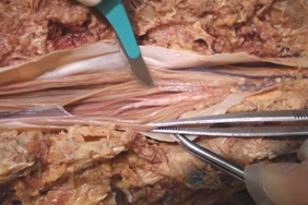
3. You can see this Youtube video for the ascending and descending tracts of the spinal cord: https://www.youtube.com/watch?v=_G-d8gKIlOw
4. Be familiar with the brain structures from each of the following pages of "Netter's Atlas of Human Neuroscience" 1st Edition (and 2nd Edition in parenthesis):
Page 24 (page 35-36): Areas 3, 1, 2, 4, 6 of Brodmann, somatosensory, motor, and premotor cortex
Page 25 (page 37, 47): Anterior commissure
Page 29 (page 38, 46, 50, 63): Denticulate ligament, dentate gyrus, gyrus fasciolaris
Page 30 (page 47): Olfactory bulb, septal nuclei, anterior nuclei of the thalamus, mammillary bodies, indusium griseum, hippocampus, amygdala, stria. terminalis, stria medullaris, habenular nuclei, interpeduncular nucleus
Page 31 (page 48): Frontal and occipital forceps of corpus callosum, medial and lateral longitudinal striae
Page 34 (page 52): Thalamus; all nuclei: midline, medial dorsal (MD), etc.; all lateral, all ventral, and intralaminar nuclei (centromedian, central lateral, parafascicular)
Page 35 (page 54-55): Pulvinar, pineal, inferior and superior colliculi, medial and lateral geniculate bodies, cerebellar peduncles, pyramid, inferior olive, choroid plexus of 4th ventricle
Page 36 (page 56-57): Cerebellum, vermis, paravermis, lingula, central lobule, culmen, declive, folium, tuber, pyramid, uvula, nodule, cerebellar nuclei
Page 38 (page 62): Filum terminale, cauda equina, conus medullaris
Page 39 (page 63): Meninges, nerve rootlets (filaments of a nerve root), and nerve roots
Page 40 (page 64): Meninges, nerve roots
Page 41 (page 65): Have a specific concept of ascending, descending, and bilateral pathways of the spinal cord
Page 44 (page 70-71): Cerebellar peduncles, locus coeruleus, sulcus limitans, tuber cinereum, colliculi, choroid plexus
Page 57-58 (page 96-97): Meninges layer
Page 87 (page 139): Dural sac (black), filum terminale, cauda equina, conus medullaris
Page 169 (page 251): Reticular formation, raphe, PAG
Page 170 (page 252): Thalamus reticular nuclei, reticular Formation
Page 174 (page 256): Cerebellum, vermis, paravermis, lingual, central lobule, culmen, declive, folium, tuber, pyramid, uvula, nodule
Page 175 (page 257): Cerebellar peduncles
Page 176 (page 260): Thalamus projections to the cerebral cortex
Page 191 (page 290): Amygdala, nucleus basalis (of Meynert)
Page 195 (page 298): Superior cerebellar peduncle
Page 206 (page 316): Locus coeruleus
Page 207 (page 317): Raphe nuclei
Page 209 (page 319): Septal nuclei, nucleus basalis (of Meynert)
Page 237 (page 353): Cerebellar peduncles
Page 243 (page 363): Corticobulbar tract
Page 244 (page 364): Corticospinal tract
Page 258 (page 380): Cerebellar nuclei: fastigial, globose, emboliform, dentate, vermis, vestibular nuclei
Page 292-293 (page 417-418): Olfactory bulb, septal nuclei, hippocampus, amygdala, stria terminalis
Page 295 (page 420): Septal nuclei
Page 298 (page 423): Anterior commissure, medial and lateral olfactory striae
Price: $950
Registration Discount: $750
You can receive the discounted price of $750 by using your CHI-Pak or by registering and making a minimum deposit of $300 at a prior CHI class and pay the balance in full 45 days prior to the class start date.
(If the class is not paid in full 45 days before the start of class, the rate automatically goes up to $950)
Repeat: $475
Thank you for the great brain course at AZCOM in January. To say it has changed my practice would be an understatement. My results are much different, and the efficacy of all my other methods is greater. I very much appreciate your contribution to my ongoing education.
We (globally, as osteopaths) are very thankful for your contribution to the body of knowledge we employ in caring for hurting people everyday. Personally, I feel as if my ability to help people has been enhanced by a quantum step/level, a "quantum leap", if you will.
David W. Asher, DO
I was speechless! I almost fell off the chair when I touched Pat’s head! I think I need to take up to Brain 14 to understand what you (Bruno Chikly) did with her. I think you stole someone else’s brain and put it in her.
Pat’s brain was supple, there was a ton of fluid in it, all the cells are moving, ALIVE, and there was no conflict in it. Part of the left brain had a lot of "conflict" before. When I sink my fingers into the cortex, there was a give. During the first session after yours, even mere intent was moving the brain. I AM NOT EXAGGERATING!
I told my staff that I am giving up my practice, family and everything and apprentice with Bruno to learn to be able to treat like that, or I will have to live up to 120 years!!
Usha Kanithi, PT
I just attended a CME program that I consider to be one of the top few educational experiences of my 30 years as an osteopathic physician. This program "Brain, Parenchyma, Nuclei and Fluid" taught by Bruno Chikly, MD, DO was offered by the Cranial Academy and your [AZCOM] OMM Department.
Paul Lee, DO, FAAO, DABMA
Your lecture and workshop were splendid…For the first time the brain component of the Primary Respiratory Mechanism was addressed as palpable structures…I was convinced that I actually felt the lateral ventricles change their orientation in your workshop.
I was amazed how quickly you located the infarction in my right temporal lobe without even knowing my stroke history.
Thank you for sharing your knowledge and experience with us.
KEEP DIGGING ON,
Fred L. Mitchell, Jr
My client who had scoliosis spinal fusion surgery and is fused from C7 to sacrum and head moved left to right and diagonally with titanium rods. The right side had tension in all three directions and the left a downward pull to the spine. After the release in Brain Therapy 2 (B2), she no longer feels the hardware pulling and the neck feels softer. She also feels more stability in her gait. Her head feels straighter and more natural. Her neck and head do not feel as locked in as before. She feels the change is very dramatic.