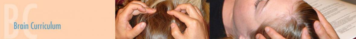
 In Brain 3 you'll deepen the skills you've already studied and have been practicing and learn how to work with and release restrictions in several new areas of the CNS. This class will add the release of all components of the peripheral body controlled by the Autonomic Nervous System (ANS): muscles, fascia, joints, organ of senses, vasculature, etc…
-
Extracellular Fluid Technique (EFT) for the periphery, spinal cord, cerebellum, and cerebrum
-
Vestibular Nuclei and Pathways (maintenance of Equilibrium: Flocculo-nodular lobe, Semicircular Canals, Utricle, Saccule)
-
Cochlear nuclei and Pathways (Auditive Pathways: anterior and posterior cochlear nuclei, cochlea, superior olive, inferior colliculi, medial geniculate, auditory cortex)
-
Functional Fulcrum for the brain
-
Three-layered cortex: Hippocampus, Dentate Gyrus, Fimbria
-
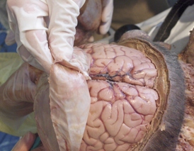
All parts of the Cerebellum: vermix, lobules and fissures
-
Three approaches to perform full CNS lesions assessment
-
Autonomic nervous system innervation: application to segments of the body, fascia, joints, viscera, diaphragm, eyes, ears, teeth, emotions, and self-treatment
-
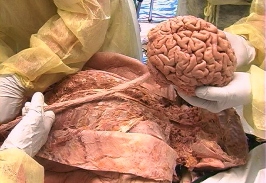
Embryologic brain and heart
-
New brain and spinal cord protocol
Class length 3 days
Conflict of Interest: All classes presented by Chikly Health Institute have no financial conflict of interest.
CHI is not sponsored by outside organizations or corporations.
Please read "Our Policies" for more information: https://chiklyinstitute.com/Policies
• Contact Continuing Education (CE) Hours Total: 18 CEUs for massage therapists - NCBTMB Approved Provider # 451238-10
NCBTMB CEUs are accepted in every US state for NCBTMB certification renewal.
Most states accept NCBTMB for license renewal but not all. We are also approved for NY state.
Please look here for more information: http://www.ncbtmb.org/map/requirements-map.
Because certification and license renewal policies vary from state to state, it's important for you to make sure the CEUs are accepted wherever you practice. Therefore, please be aware that this information may not apply in your state.
Check your state’s website at: http://www.ncbtmb.org/regulators/state-info.
Alberta massage therapists: Members of the RMTA will receive 15 Continuing Education Credits (CEC) upon the submission of a certificate of completion for each course.
• 18 hours approved by the Massage Therapy Association of Manitoba, Canada (MTAM)
• 18 hours approved by the Certified Registered Massage Therapy Association of Alberta, Canada (CRMTA)
We are in the process of providing Continuing education for numerous other professions. Please check back to this page later as we will post all updates.
-
Extracellular Fluid Technique (EFT) for the periphery, spinal cord, cerebellum, and cerebrum
-
Vestibular Nuclei and Pathways (maintenance of Equilibrium: Flocculo-nodular lobe, Semicircular Canals, Utricle, Saccule)
-
Cochlear nuclei and Pathways (Auditive Pathways: anterior and posterior cochlear nuclei, cochlea, superior olive, inferior colliculi, medial geniculate, auditory cortex)
-
Functional Fulcrum for the brain
-
Three-layered cortex: Hippocampus, Dentate Gyrus, Fimbria
-
All parts of the Cerebellum: vermix, lobules and fissures
-
Three approaches to perform full CNS lesions assessment
-
Autonomic nervous system innervation: application to segments of the body, fascia, joints, viscera, diaphragm, eyes, ears, teeth, emotions, and self-treatment
-
Embryologic brain and heart
-
New brain and spinal cord protocol
COURSE SCHEDULE (Subject to change)
Day One:
8:30 Registration
9:00 - 11:00 Introduction, teachers, students, teaching assistants, and facilitator. Teaching material
Embryological connection Head-Heart
The extracellular fluid technique (EFT) for the brain
11:00 - 11:15 Break / group discussion
11:15 - 12:30 Vestibular nuclei and pathways
12:30 - 2:00 Lunch
2:00 - 3:30 Cochlear nuclei and pathways
3:30 - 3:45 Break / group discussion
3:45 - 5:30 Functional Fulcrum of the Brain
Day Two:
9:00 - 11:00 Questions and answers
Three-layered cortex: cerebellum
11:00 - 11:15 Break / group discussion
11:15 - 12:30 Three-layered cortex: hippocampus part 1
12:30 - 2:00 Lunch
2:00 - 3:00 Three-layered cortex: hippocampus part 2
3:00 - 3:30 Three-layered cortex: dentate gyrus
3:30 - 3:45 Break / group discussion
3:45 - 4:45 Finding dominant lesions in the CNS: fascia and fluid approaches.
4:45 – 5:30 Organization of the Autonomic Nervous System (ANS) system part 1
ANS technique applied to articulations
Day Three:
9:00 - 10:30 Questions and answers
Organization of the ANS system part 2.
Case studies.
ANS technique applied to the fascia
10:30 - 10:45 Break / group discussion
10: 45 - 12:45 ANS technique applied to viscera
12:45 - 2:00 Lunch
2:00 - 3:30 ANS technique applied to Self-Treatment
Take home Protocol.
Final questions and answers.
Self-Reflection and identification of changes for practitioner’s practice.
LEARNER OBJECTIVES (Subject to change)
- By the end of the 1st-day participants will be able to correctly demonstrate on a live person Brain 3 Extracellular fluid technique (EFT) for the brain
- By the end of the course participants will be able to correctly demonstrate on a live person how to release dysfunction of the Vestibular nuclei
- By the end of the course participants will be able to correctly demonstrate on a live person how to release dysfunction of the Cochlear nuclei
- By the end of the course participants will be able to correctly demonstrate on a live person how to release the Brain using Functional Fulcrum.
- By the end of the course participants will be able to correctly demonstrate on a live person how to release dysfunction of the cerebellum
- By the end of the course participants will be able to correctly demonstrate on a live person how to release dysfunction of the hippocampus
- By the end of the course participants will be able to correctly demonstrate on a live person how to release dysfunction of the dentate gyrus
- Based on a fascia and fluid approach participants will be able by the end of the course to correctly find on a live person a dominant dysfunction of the CNS
- By the end of the course participants will be able to correctly demonstrate on a live person how to use Brain 3 ANS technique to release articular dysfunction
- By the end of the course participants will be able to correctly demonstrate on a live person how to use Brain 3 ANS technique to release dysfunction of the fascia
- By the end of the course participants will be able to correctly demonstrate on a live person how to use Brain 3 ANS technique to release dysfunction of the viscera
- By the end of the course participants will be able to correctly demonstrate on themselves how to use Brain 3 ANS technique to release dysfunction of the joints, fascia or viscera
INSTRUCTIONAL METHODS
- Lecture
- Study Guide
- Question & Answer
- PowerPoint Slides
- Demonstration
- Practice sessions
- Review
B2 plus at least four (4) months of practice after attending B2.
An in-depth advanced study of anatomical terms and concepts is necessary; a study list is provided below.
Anatomical/Physiological Terms
The DVD "Dissection of the Brain" and Spinal Cord is a great preparation for the Brain 3 class. Please pay special attention to chapter 13 "cerebellum" to facilitate your experience.
Be sure you understand the following words and, as applicable, know precisely where these structures are located in the body. For this class, it is more important to know their 3-dimensional location and relationship with one another, than their classically described physiology.

We will go over the vestibular and cochlear systems, the three-layer cortex (hippocampus and cerebellum)
Be aware of the complex subdivisions of the cerebellum:
-
THREE LOBES: Anterior Lobe, Posterior Lobe, Flocculo-Nodular Lobe.
-
VERMIS (LLCC): Lingula, Central Lobule (Lobulus Centralis), Culmen, Declive, Folium, Tuber, Pyramis, Uvula, Nodule.
-
LOBULES & FISSURES: Quadrangular (Superior Quadrangular) Lobule, Simplex (Inferior Quadrangular) Lobule, Posterior Superior Fissure - Superior Semilunar (or Crus I or Ansiform) Lobule, Horizontal Fissure, Inferior Semilunar (or Crus II or Ansiform) Lobule, Gracilis Lobule, Biventral Lobule, Secondary Fissure (Retrotonsillar), Tonsils of the Cerebellum
Superior olive, inferior colliculi, medial geniculate, hippocampus layers, dentate gyrus, fimbria, part of the cerebellum, autonomic innervation, sympathetic and parasympathetic nervous system, pre and post-ganglionic (synaptic) neurons, enteric nervous system, sympathetic ganglia, intermediolateral cell column, paravertebral ganglion, superior, middle and inferior cervical ganglion, stellate ganglion, sympathetic Plexi, celiac plexus, superior and inferior mesenteric ganglia, aorticorenal ganglia, greater splanchnic nerves.
Please read the autonomic nervous system text below, especially the organization of the sympathetic system.
Be familiar with the brain structures from each of the following pages of "Netter's Atlas of Human Neuroscience" 1st Edition (2nd Edition in parenthesis):
Page 30 (page 47): Hippocampus, dentate gyrus
Page 32 (page 50): Hippocampus, fimbria
Page 35 (page 54-55): Posterolateral View: Pineal, superior and inferior colliculi, medial and lateral geniculate, Pulvinar, olive, vestibular area
Page 36 (page 56-57): It would be very helpful to study every word related to the cerebellum found on these pages.
Page 116 to 120 (page 172-176): Autonomic innervations
Page 121-122 (page 177-178): Cervical sympathetic ganglia
Page 126 (page 185): Sympathetic innervation, stellate ganglion
Page 129 (page 188): Sympathetic Plexi, celiac plexus, superior and inferior mesenteric ganglia, aorticorenal ganglia, hypogastric Plexi
Page 132 (page 195): Enteric nervous system
Page 136 (page 201): Autonomic innervations
Page 141 (page 208): intermediolateral cell column
Page 152-153 (page 224-227): Vestibular and cochlear nuclei
Page 158 (page 235-236): Vestibular and cochlear nuclei
Page 163 (page 245): Vestibular and cochlear nuclei
Page 173 (page 255): Cerebellar lobes and regions
Page 174 (page 256): All divisions of vermix
Page 224-225 (page 336-337): Vestibular and cochlear system
Page 226 to 229 (page 338-342): Cochlear cochlear system
Page 230-231 (page 343-344): Vestibular system and pathways
Page 247 (page 368-369): Vestibular pathways
Page 253 (page 375): Cerebellar regions
Page 254 (page 376): Three layers of the cerebellum
Page 257-258 (page 379-380): Vestibular and cochlear nuclei
Page 289-290 (page 414-415): Hippocampus, dentate gyrus, fimbria
Some resources
-
"Netter's Atlas of Human Neuroscience", Icon Learning System
-
"Atlas of Anatomy", Thieme, Head and Neuroanatomy, ISBN: 978-1-58890-441-6
-
"Color Atlas of Human Anatomy, Vol. 3, Nervous System and Sensory Organs", Thieme, ISBN: 978-1-58890-0647
-
"Neuroanatomy, 3-D Stereoscopic Atlas", M. Hirsch, T. Kramer, Springer Ed, ISBN: 3-540-65998-6
-
"The Human Brain", John Nolte, Mosby, ISBN: 978-0-323-01320-8
-
"Neuroanatomy, Text and Atlas", John Martin, Appleton & Lange Ed ISBN: 0-8385-6694-4
-
Autonomic Nervous System: Review
It is important to study the information below and have an appreciation of the Somatic and Autonomic (Vegetative) Nervous System
I. The Somatic and Autonomic Nervous System (ANS)
The somatic nervous system is a voluntary system that allows us to consciously interact with our environment. It has one simple action: causing contraction of skeletal muscles. It has one fast, myelinated motor neuron outside of the central nervous system (CNS) and no ganglia. It has one neurotransmitter: acetylcholine, always activating its effector.
The autonomic nervous system (ANS) is a system of information not under direct voluntary control. The word "autonomic" comes from two Greek words that mean "self" and "law".
The ANS allows the body to respond involuntarily, subconsciously, and automatically to the body's demands for daily adjustments and maintenance of homeostasis/allostasis.
The ANS has 2 simple, mainly involuntary actions to maintain the internal environment:
Contraction of smooth muscles (viscera, eyes, blood vessels, etc.) and cardiac muscles
Glandular secretion (adrenal, lacrimal, salivary, digestive, cutaneous, etc.)
The ANS is also called the general visceral motor system.
The information received by the ANS primarily comes from the surface of the body (somatic sensory), the viscera (visceral sensory), and the external environment (special sensory). The amount of sensory receptors in the viscera is 10% of the number of receptors of the skin.
Two divisions have traditionally been associated with the ANS: the parasympathetic and sympathetic nervous systems. They harmonize and counteract each other. Under normal conditions the two main branches of the ANS are usually in balance.
A third division, the *benteric nervous system*p, deals with the diffuse visceral (especially enteric) sensory input, has more recently been isolated (Michael Gershon, Columbia University).
As we saw previously, the somatic nervous system is located outside of the central nervous system (CNS). It is mainly a one-neuron system outside of the CNS (synapse spinal cord-voluntary muscle).
The ANS is comprised of at least 2 systems (sympathetic and parasympathetic) and 2 neurons outside of the central nervous system that synapses together.
The 2 neurons outside of the CNS are called the pre and postganglionic neurons. This added interneuron gives the ANS its autonomy and capacity to make local "decisions". Our practice will be to localize and work on these groups of local/regional/central control centers (GCC).
We will call the first group of ANS control centers (GCC) group "0". GCC 0 is a CNS grouping of cortical and subcortical centers responsible for the central control of the ANS.
The GCC 0 is comprised of:
-
The reticular formation
-
The hypothalamus
-
The thalamus
-
The pituitary
-
The prefrontal cortex
-
The orbital cortex
-
The cingulate gyrus
-
The olfactory cortex
-
Numerous brainstem nuclei, etc.
ANS main hypothalamic nuclei centers:
-
Paraventricular
-
Anterior
-
Posterior
-
Lateral
ANS main thalamic nuclei centers:
-
Mediodorsal (MD)
-
Medioventral (MV)
-
Centromedian (internal medullary lamina)
The traditional description of the system is that preganglionic sympathetic and parasympathetic neurons use acetylcholine (ACh) type nicotinamic as their main neurotransmitter.
Postganglionic sympathetic neurons use mainly adrenaline/noradrenaline (NA). Postganglionic parasympathetic neurons use mainly acetylcholine type muscarinic (M). Stomach acid secretion is mediated by M1, cardiac manifestations by M2; most other visceral parasympathetic neurons are mediated by M3 receptors.
These simple notions are been slowly replaced by the concept of co-transmitters (multiple neurotransmitters in one synapse) and neuromodulators (they are released, but have no direct action on the postjunctional receptors). Such substances include dopamine, ATP, nitric oxide, Peptide Y, VIP, substance P, etc.
Most organs have a dual innervation (sympathetic and parasympathetic), the main exceptions being:
1. Organs with only parasympathetic nervous system innervation:
The bronchi (no sympathetic innervation, but the adrenaline in the bloodstream can have a similar effect)
The lacrimal gland (no sympathetic innervation)
2. Organs with only sympathetic nervous system (SNS) innervation:
The blood vessels. All arteries and veins of any size have an exclusive sympathetic innervation
The adrenal medulla (epinephrine/adrenaline)
The sweat glands (acetylcholine)
The hair follicles
Note that superior limbs, inferior limbs, and body walls have no direct parasympathetic innervation.
II. Embryology of the ANS
All of the ANS derive embryologically from the neural crests.
Neural crests are structures located on the superior lateral aspect of the neural tube. Neural crest differentiation began mainly in the cranial region at the stage of the neural fold. They have been referred to as the fourth germ layer. Later on, three regions of the neural tube generated neural crests: rhombencephalon, mesencephalon, and prosencephalon, each with a different migratory pattern. They migrated throughout the embryo acting as agents bearing information that would modify target tissues and form many of the structures of the embryo. Numerous structures are derived from neural crests, such as:
CNS:
-
Cranial nerve ganglia
-
All associated glial cells
-
Dura from mesoderm
-
Pia and arachnoid mater
ANS:
-
Sympathetic ganglia
-
Sympathetic plexuses: celiac, mesenteric, renal
-
Adrenal medulla (chromaffin cells)
-
Parasympathetic ganglia
PNS:
-
Sensory (dorsal root) ganglia
Schwann cells
Other:
-
Head mesenchyme
-
Face
-
Most of the ocular globe
-
Pharyngeal arches
-
The connective tissue around the eye
-
C cells of the thyroid gland
-
Cartilage rudiments (nose, face, middle ear)
-
Dermis
-
Melanocytes
-
Smooth muscle and adipose tissue
-
Odontoblast of the teeth
III. Structure and Function of the ANS
Parasympathetic Function - "The Craniosacral Division": "Rest & Digest"
The anabolic system, the parasympathetic system conserves and helps restore body energy resources. It helps regenerate injured tissues and stimulates immune functions.
It is generally most active during sleep and deep relaxation states.
Each individual parasympathetic pathway is regulated independently. The ratio of preganglionic to postganglionic neurons is 1:1 (except vagus nerve).
Parasympathetic activation is more local than sympathetic activation.
A. Effects of parasympathetic stimulation:
-
Decreases heart rate (M2)
-
Decreases respiratory rate
-
Decreases blood pressure
-
Increases blood flow to the skin
-
Increases gastrointestinal motility, kidney function (M3)
-
Increases secretion of lacrimal glands (M3)
-
Increases salivary glands, watery secretion (M3)
-
Increases digestive glands
-
Increases gastric acid secretion (M1)
-
Increases bronchial glands
-
Constricts bronchioles (M3)
-
Relaxes sphincters (M3)
-
Contracts pupils (miosis)
-
Contracts ciliary muscles (accommodation) (M3)
-
Contracts urinary bladder (M3)
-
Stimulates erection (M3)
No parasympathetic innervation of the limbs, body wall, blood vessels (except a possible innervation of the coronary arteries of the heart), the sweat glands, the hair follicles, and the adrenal medulla.
B. Anatomy of the Parasympathetic System:
The parasympathetic system innervates the cranium, neck, thorax, and abdomen, but not the extremities.
Preganglionic parasympathetic nerves are very long and are thinly myelinated (peripheral nerve type B).
-
Pre-synaptic neurons: hypothalamus, brainstem, and sacral segments of the spine.
-
Pre-synaptic neurotransmitter: Acetylcholine
-
Pre-ganglionic neurons are in cranial nerves III, VII, IX, X and in the lateral horn of sacral segments S2, S3, S4 (splanchnic nerves)
Postganglionic nerves are typically short and are unmyelinated (type C or IV). Fibers usually synapse close to or within the organ they innervate. They can use the same nerve plexus as the sympathetic system.
Usually, the ratio of preganglionic to postganglionic fibers is 1:1 (except for the vagus nerve). The parasympathetic system has little "divergence".
C. Specific Anatomy of the Parasympathetic System:
1. Cranial Division
The cranial nerves nuclei include:
Edinger-Westphal nucleus (III)
Superior salivatory nucleus (VII)
Inferior salivatory nucleus (IX)
Two nuclei of the vagus (X): dorsal motor nucleus and nucleus ambiguus
Parasympathetic ganglia associated with the cranial division:
Edinger-Westphal nucleus (III): ciliary ganglion for papillary constrictor muscle (iris) and ciliary muscle (lens)
Superior salivatory nucleus (VII): pterygopalatine ganglion for lacrimal and nasal glands;
Submandibular ganglion for submandibular and sublingual salivary glands
Inferior salivatory nucleus (IX): otic ganglion for the parotid gland
Two nuclei of the vagus (X):
-
Dorsal motor nucleus (thorax + abdomen)
-
Nucleus ambiguus:
dorsal division: palate, larynx, esophagus
ventrolateral division: heart
The vagus (cranial nerve X), one of the most important parts of the parasympathetic nervous system is a nerve approximately 80% sensory (tractus solitarius) and 20% motor. It innervates both somatic and visceral organs in the neck, chest, and abdomen.
2. Sacral Division
Sacral segment S2-S4 (intermediolateral spinal cord segment S2-S3-S4) Innervate: left 1/3 transverse colon, descending colon, rectum, bladder, reproductive organs
2. Sympathetic Function - The "Thoracolumbar Division": "Fight or Flight"
The catabolic system, the sympathetic system expends body energy resources, depresses immune functions, and releases epinephrine (adrenaline) and norepinephrine (noradrenaline) hormones.
The Sympathetic Nervous System is often activated as a whole.
A single preganglionic axon contacts many cells. Postganglionic axons can receive input from approximately 30 different preganglionic fibers (important) "divergence"). Sympathetic activation is usually a diffuse activation with multiple systems activated concurrently.
We are going to work mainly on the sympathetic system in this class because:
1. The sympathetic NS has more general effects (rather than local). It has more widespread effects, usually mass responses (more divergence).
2. Is more distributed to the whole body.
3. Has effects lasting relatively longer: it stimulates the medulla of adrenal glands that secrete hormones in the blood circulation.
But let's remember the parasympathetic nerves have about 3 times more sensory axons (sensations of fullness, tension, etc.), than sympathetic (usually pain messages).
Sympathetic functions are globally the reverse of parasympathetic functions.
A. Effects of sympathetic stimulation
-
Increases heart rate (beta á1)
-
Increases blood pressure
-
Vasoconstricts blood vessel (alpha 1)
-
Increases respiratory rate
-
Decreases blood flow to the skin
-
Increases secretion of epinephrine
-
Increases secretion of sweat glands (acetylcholine)
-
Piloerect hairs
-
Increases blood sugar level
-
Decreases gastrointestinal motility, kidney functioning
-
Decreases secretion of digestive glands
-
Constricts sphincters; causes spasm
-
Dilates the pupils (mydriasis) (T1, via carotid)
-
Stimulate fat cells via Beta 3 receptors and induces lipolysis
-
Stimulates ejaculation
Pain is usually carried by sympathetic (or somatic) nerves. No sympathetic innervation of the bronchi and the lacrimal glands.
B. General Anatomy of the Sympathetic System:
Sympathetic fibers leave the spinal cord (white ramus) and usually synapse in one ganglion of the paravertebral sympathetic chain.
Most preganglionic sympathetic nerves are short and are myelinated, whereas the postganglionic nerves are long and are unmyelinated.
Each preganglionic cell gives rise to multiple fibers that innervate cells in either the paravertebral sympathetic chain, the prevertebral ganglia (i.e. celiac, aorticorenal and mesenteric), or the few terminal ganglia in the target organs (i.e. adrenal gland). Usually, the ratio of preganglionic to postganglionic fibers is one to 15 or 30.
Preganglionic neurons (acetylcholine) are located in the lateral horn of T1 to L2 spinal cord segments. They innervate most of the body. Postganglionic neurons: release noradrenaline (NA) onto Alpha-1, Beta-1, Beta-2, or Beta-3 adrenergic receptors.
Exceptions, the following sympathetic structures don't release norepinephrine:
Adrenal medullary chromaffin cells secrete epinephrine
Sympathetic nerves innervating sweat glands secrete acetylcholine
Sympathetic nerves innervating blood vessels in skeletal muscle secrete acetylcholine
In some animals, there are also sympathetic cholinergic fibers that go to blood vessels supplying muscle
Muscle
Sympathetic nerves innervating renal blood vessels secrete dopamine
The group of the control center 1 (GCC 1) is the intermediolateral segments of the spinal cord: T1 to L2 or L3.
The group of the control center 2 (GCC 2) is the sympathetic chain (paravertebral): cervical, thoracic, lumbar, and sacral ganglia and 1 midline impar (coccygeal) ganglion.
Leaving only the ganglion, sympathetic neurons can bifurcate medially and innervate the viscera, laterally for the body wall (blood vessels, sweat glands, arrectors pilorum muscles) or go up or down the sympathetic chain.
Only 13 or 14 ganglia receive direct sympathetic branches (white rami) from the spinal nerve, the rest have only grey (unmyelinated) communicans, receiving nerves ascending or descending from the sympathetic chain.
Sympathetic distribution:
1. The synapse of the sympathetic chain that innervates:
-
All the blood vessels
-
Arrector pili muscles
-
And sweat glands
-
Always in the paravertebral chain
2. The synapse of the sympathetic chain innervating the head and neck is always in the paravertebral cervical chain (one of the 3 sympathetic cervical ganglia)
3. The synapse of the sympathetic chain innervating: the thorax and limbs are always in the paravertebral chain: C1-T5
4. The synapse of the sympathetic chain innervating the abdomen and pelvis is always in the preaortic (prevertebral) ganglia. This is the 3rd group of control center (GCC 3): celiac ganglion, superior mesenteric ganglion, aorticorenal ganglion, and inferior mesenteric ganglion. The postganglionic neuron will usually synapse again to a neuron of the enteric nervous system (and not directly in the organ tissue).
C. Specific Anatomy of the Sympathetic System:
GCC 2: sympathetic chain (paravertebral). There are usually 22 to 23 pairs of paravertebral ganglia: cervical, thoracic, lumbar, and sacral ganglia, and one midline impar (coccygeal) ganglion.
1. Principal Cervical Paravertebral Ganglia
Three cervical ganglia in front of cervical transverse processes:
-
Superior cervical ganglion
-
Middle cervical ganglion
-
Inferior cervical ganglion or Stellate ganglion
A. Superior Cervical Ganglion:
Also called the ganglion of Hirschfeld, it is the largest cervical ganglion (4-5 cm in length, 1 cm in width) and is located behind the internal carotid at the level of the transverse process of C2-C3 (from C1 to the angle of the mandible). Posterior to it is the longus capitis muscle. This ganglion is usually made up of the agglomeration of four ganglia, C1 to C4.
The superior cervical ganglion mainly supplies the head and neck and the heart. It has many branches including the pharyngeal nerves, laryngeal, and superior cardiac nerve. It runs superiorly to the carotid canal, "hitchhiking" along the carotid arteries and their branches, connecting with a carotid ganglion, then further to the carotid plexus, the cavernous sinus where it meets branches from cranial nerve III, IV, optic ganglion of V, VI, Tympanic nerve of IX.
B. Middle Cervical Ganglion or Thyroid Ganglion:
The smallest of the three cervical ganglia, sometimes not present. It is located at the level of the transverse process of C6 or C7. It is usually superior or close to the inferior thyroid artery. It is the coalescence of the ganglion of C5 and C6. The middle cervical ganglion mainly supplies the area of the neck and the heart. It branches into the middle cardiac nerve, the largest of the three cardiac nerves.
C. Inferior Cervical Ganglion or Stellate Ganglion:
It is about 0.8 to 1 cm in length and lies superior to the apex of the lung. It is anterior to the transverse process of C7 and the 1st rib. It is often the fusion of the ganglion of C7 with the first thoracic ganglion. It is at the level of the transverse process of C7 or T1.
The inferior cervical ganglion mainly supplies the posterior cranial arteries, lower neck, upper extremities, and heart. It branches into the inferior cardiac nerve. The left stellate ganglion is lower than the right.
2. Thoracic and Abdominal Ganglia: Paravertebral Ganglia
Ten to twelve dorsal paravertebral ganglia: mostly in front of the head of the ribs, covered by pleura (except dorsal 11 and 12, lateral to the body of vertebrae). The nerves have to pierce the diaphragm.
Prevertebral ganglia:
The 3rd group of control centers (GCC 3) is the prevertebral (preaortic) ganglia.
Principal prevertebral ganglia:
Celiac ganglion or semi lunar ganglion ("solar plexus", epigastric plexus)
Largest ganglion in the body. It is anterior to the aorta and the crura of the diaphragm.
Superior mesenteric ganglion
Inferior mesenteric ganglion
Aorticorenal ganglion
The medulla of the adrenal glands is sometimes compared to modified sympathetic ganglia (postganglionic neurons) because there are only preganglionic fibers (T8-L1) stimulating them - no postganglionic fibers. The adrenals (suprarenal gland) weigh about 8-10 g, anterior to the body T12-L1 and rib 11 and 12th.
Adrenal glands are made of a capsule, an outside cortex (with 3 layers, zona glomerulosa for aldosterone, zona fascicularis, and zona reticularis for cortisol and sexual hormones), and the medulla in the center for the catecholamines: epinephrine (adrenaline) and norepinephrine (noradrenaline).
The medulla of the adrenal glands is also part of GCC 3.
The chromaffin cells in the medulla, the center of the adrenal glands, secrete the catecholamines: epinephrine (adrenaline) and norepinephrine (noradrenaline).
The greater splanchnic nerve (T5-T10) innervate the medulla of the adrenal glands and stimulate directly (preganglionic fibers) its chromatofinn cells that release approximately 80% of adrenaline and 20% of noradrenaline into the blood. Note that this percentage is inversed for the CNS, where the locus ceruleus releases 80% of noradrenaline.
The medulla of the adrenal glands is the only place where the body can convert norepinephrine to epinephrine. Epinephrine is secreted into the bloodstream, so its effects are similar to norepinephrine. Epinephrine effects last about 10 times longer.
Catecholamine production:
Tyrosine convert to Dopa
Dopa to Dopamine
Dopamine to norepinephrine, then to epinephrine in the adrenal glands
3. Lumbo-Pelvic Ganglia: Paravertebral Ganglia
In the abdomen and pelvis, prevertebral ganglia are near the ventral surface of the vertebral column.
-
Four lumbar ganglia anterior or lateral to the body of the vertebrae
-
Three to five sacral ganglia and the impar (meaning unpaired) or coccygeal ganglion, all in front of the sacrum, medial to the anterior sacral foramina. The impar ganglion is usually 3-4 mm in diameter, located anterior to the last coccygeal vertebrae. It may have a nervous and a neurocrine function.
Some fibers going to the abdominopelvic area can synapse in a prevertebral ganglion located close to the abdominal aorta or branches (splanchnic nerves).
The splanchnic nerves are presynaptic nerves, having a synapse in the GCC 3 that contribute to visceral innervation.
Main splanchnic nerves:
-
Greater splanchnic nerve-spinal level origin: T5 through T8, T9, or T10 (to celiac ganglia)
-
Lesser splanchnic nerve (94%)-spinal level origin: T9 through T10 or T11 (to superior
-
Mesenteric ganglia)
-
Lower (least) splanchnic nerve (56%)-spinal level origin: T12 (to renal ganglia)
-
Sacral splanchnic nerves
The ganglia and fibers make a number of ANS plexi:
-
Cardiac plexus
-
Pulmonary plexus
-
Diaphragmatic plexus
-
Celiac plexus
-
Renal plexus and superior Mesenteric plexus
-
Adrenal plexus
-
Inferior Mesenteric plexus
-
Hypogastric (pelvic) plexus
-
Ovarian or spermatic plexus
The 4th group of control centers (GCC 4) could be almost anywhere (anatomical variations). Usually close to the known ganglion, or close to arteries or groups of lymph nodes (high innervation)
3. The Diffuse Visceral (Enteric) Nervous System
All viscera are innervated by the sympathetic and parasympathetic systems.
The enteric nervous system includes as many as one billion neurons, 1/100th of the number of neurons in the brain, and much more than the number of neurons in the spinal cord.
There are two main layers of neurons throughout the wall of the intestines and act independently of sympathetic and parasympathetic innervation:
-
External Plexus: Myenteric Plexus of Auerbach. It is located between the inner circular muscle layer and the outer longitudinal muscle layer. This plexus is responsible for most smooth muscles movements (peristalsis) of the intestines.
-
Inner Plexus: Submucosal Plexus of Meissner (absent in the stomach and esophagus). It is responsible for sensing the environment of the intestines and regulating its response (vascularization, cell metabolism, ion, and water transport).
Depending on conditions, they can be either excitatory or inhibitory.
There are numerous glial cells in the enteric nervous system, as well as the interstitial cell of Cajal (ICC). This cell is not neural or glial, but somehow in between, it acts as the intrinsic pacemaker of the bowel.
REVIEW
-
GCC 0: CNS cortical and subcortical centers including the reticular formation, hypothalamus, pituitary, etc.
-
GCC 1: The intermediolateral segments of the spinal cord: T1 to L2 or L3.
-
GCC 2: Sympathetic chain (paravertebral). 22 to 23 pairs of paravertebral ganglia: cervical, thoracic, lumbar, and sacral ganglia, and 1 midline impar (coccygeal) ganglion.
-
GCC 3: Preaortic (prevertebral) ganglia: celiac ganglion, superior mesenteric ganglion, aorticorenal ganglion, inferior mesenteric ganglion + adrenal medulla.
-
GCC 4: They could be almost anywhere (anatomical variations). Usually close to the known ganglion, or close to arteries or groups of lymph nodes (high innervation).
Price: $950
Registration Discount: $750
You can receive the discounted price of $750 by using your CHI-Pak or by registering and making a minimum deposit of $200 at a prior CHI class and pay the balance in full 45 days before the class start date.
(If the class is not paid in full 45 days before the start of class, the rate automatically goes up to $950)
Repeat: $475
I would like to thank you so much for the brain treatment. It has been gradually taking effect over the past week and a half. I have noticed distinct improvements in memory recall, calculation, and thinking speed. I feel the best I have in quite a few years. Since my bike accident I have felt quite flat - neither happy or really sad - so having my sense of humor back is also a big change...it is the best help I've had with the cognitive and emotional injuries since at least 7 years ago.
Scott C, DO
The class was great. Just to think that you could located, feel and then treat such specific parts of the brain was wonderful. Everyone who works with individuals who present with any type of brain damage should take this class. I have used it for all of the children that I work with and it has produced some fantastic changes in a short period of time. I highly recommend B1 for all practitioners.
Mable Sharp
This is the best class I ever take in 20 years taking all sorts of hands-on seminars.
Robin Walpert
This is an extraordinary class. Dr Chikly has developed a profound method of releasing trauma from tissue anywhere in the body (including brain trauma). The technique is subtle, gentle and very harmonizing for the client.
Robert H. Weiner
This course is so important to me that I repeated it in December ’06.
The content is key to healing and Dr. Chikly is key in explaining and demonstrating it.
Dr. Chikly has a special way of communicating the curriculum which is precise and professional while at the same time humorous and personal.
Alaya Chikly is a special and essential addition to the class, providing guided meditations and other less traditional exercises which varied the presentation and opened the minds of the students to new and more expansive ways of perceiving this unique and useful material.
There is no question in my mind that this work is an important part of what I do daily with the people who come to see me for help with a wide variety of health issues. There is no way I would ever give up what I have learned from Dr. Chikly, nor could I envision my practice without it. I am deeply indebted to the Chikly’s for their skill as pedagogs and their care and precision as research scientists. I plan to continue with B2 and future courses which the Chikly’s develop
Jane Shepard, LMT
B1 was one of the best courses I assisted in my last 17 years. Dr. Chikly covered thoroughly both the structural and the functional anatomy of the brain. It was a solid, precise and fascinating information. I consider this class indispensable to practitioners of any advanced modality. It should be a pre-requirement to high level therapists training. I’m using the many tools learned in B1 in almost all my patients, and being delighted with the results. The functional modifications of the brain are happening at deeper level and faster pace. The techniques learned in B1 classes became true blessings to my little handicapped patients.
Shanna Ringel, DOM, CST-D
I just attended a CME program that I consider to be one of the top few educational experiences of my 30 years as an osteopathic physician. This program "Brain, Parenchyma, Nuclei and Fluid" taught by Bruno Chikly, MD, DO was offered by the Cranial Academy and your [AZCOM] OMM Department.