Class Hours:
Registration: 8:30 a.m. First day
Class: 9:00 a.m. - 5:00 p.m.
Last Day: 8:00 a.m. - 3:30 p.m.
Times are subject to change.
Class Description:
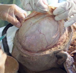
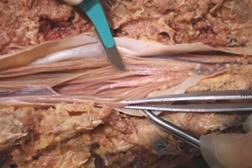
Brain 2 teaches the form, function, and response mechanisms of the brain's various components and offers hands-on techniques to effectively release many primary restrictions that can affect the whole body. In this class, we will learn different ways to find a primary lesion in the brain, and we will focus a little more on the spinal cord's grey and white matter.
Attire/Supplies:
Bring comfortable, loose-fitting clothes. Some of these approaches require touching exposed skin. We encourage you to bring a bath towel, lab clothes, and appropriate undergarments to be comfortable.
Short fingernails are required for some techniques.
Due to the potential chemical sensitivities of your classmates, please refrain from wearing perfumes or oils to class.
Please let the institute know if you can bring a treatment table.
Cancellation Policy:
Tuition Transfer: The tuition is fully transferable up to 30 days before the start of the class.
Within 14 days, a $200 administrative fee will apply.
Within seven (7) days, there is no refund of the deposit.
Tuition Refund: Tuition refund requests must be made in writing. Emails are acceptable. Tuition is fully refundable up to 30 days before the start of the class, after which your tuition is non-refundable. In case of an emergency, any requests for a refund later than 30 days before class starts will be considered on a case-by-case basis.
Seats are limited. Reserve today: : 480-999-0808 or [email protected]
For any other inquiries, contact [email protected]
193 Drury Inn & Suites Columbus Polaris
8805 Orion Place
Columbus, OH 43240
(614) 854 0216
druryhotels.com
193 Drury Inn & Suites Columbus Polaris
8805 Orion Place
Columbus, OH 43240
(614) 854 0216
druryhotels.com
https://www.druryhotels.com/bookandstay/newreservation/?groupno=10157277
Please make your reservations by Sunday, March 8, 2026 to receive your group rate. Reservations made after this date
will be subject to prevailing rates and availability. Reservations may also be made by calling 1-800-325-0720 and referring
to your group number 10157277.
At Drury Hotels, we know you have enough to worry about when traveling. In addition to the great rate, our generous
amenities will brighten your group’s day and make your journey easier.
Complimentary Hot Breakfast - Enjoy scrambled eggs, oatmeal, fresh fruit, potatoes, pastries, the all-
important coffee and more. Hot, fresh, and, best of all, free. Free hot breakfast is served daily from 6-
9:30 a.m. on weekdays and 7-10 a.m. on weekends.
Complimentary Evening Drinks and Snacks* - Join us from 5:30 p.m. – 7:00 p.m. every evening to
enjoy complimentary hot food and cold beverages at our 5:30 Kickback®. The Hotel features a rotating
menu of hot food, beer, wine, mixed drinks and soft drinks.
Complimentary Wi-Fi Throughout the Hotel - Get the score, check social networks or email family
members in the hotel!
On-Site Facilities - Take advantage of the business center, fitness center or pool while away from
home. Print boarding passes, finish a presentation or check e-mail in the Hotel’s business centers.
Anatomical/Physiological Terms
1. The DVD "Dissection of the Brain" and Spinal Cord is a great preparation for the Brain 2 class.
2. Be sure you understand the following words and, as applicable, know precisely where these structures are located in the body.
For this class, it is more important to know their 3-dimensional location and relationship with one another, than their classically described physiology.
- Reticular alarm system: median (raphe nuclei), medial and lateral columns, locus ceruleus, sulcus limitans, rhomboid fossa.
Frontal and occipital forceps of the corpus callosum
Internal capsule
Anterior commissure, medial and lateral olfactory striae, posterior commissure
The commissure of the fornix (psalterium or lyre of David)
Mamillothalamic tract
Posterior limb of the internal capsule, pyramidal decussation, corticospinal tracts
Dentate gyrus, gyrus fasciolaris
All nuclei of the thalamus, including midline, medial dorsal (MD), all lateral and all ventral, intralaminar nuclei (centromedian, central lateral, parafascicular), etc.
Nucleus accumbens, septal nuclei, nucleus basalis (of Meynert), ventral tegmental area (VTA), stria terminalis, stria medullaris
Habenular nuclei
Vermis, superior, middle, and inferior cerebellar peduncles
Vestibular nuclei
Periaqueductal gray matter (PAG)
Dura, arachnoid, pia, in the brain and spine
The spinal cord, foramen magnum, C1, C2, cervical and lumbar enlargements, dorsal and ventral roots, dorsal root (spinal) ganglion, conus medullaris, cauda equina, filum terminale
It will be helpful to read the article "CSF and Lymph: Is Human CSF Reabsorbed by Lymph?" from "Silent Waves, Theory & Practice of Lymph Drainage Therapy", Part 5, Chapter 8.
3. You can see this Youtube video for the ascending and descending tracts of the spinal cord: https://www.youtube.com/watch?v=_G-d8gKIlOw
4. Be familiar with the brain structures from each of the following pages of "Netter's Atlas of Human Neuroscience" 1st Edition (and 2nd Edition in parenthesis):
Page 24 (page 35-36): Areas 3, 1, 2, 4, 6 of Brodmann, somatosensory, motor, and premotor cortex
Page 25 (page 37, 47): Anterior commissure
Page 29 (page 38, 46, 50, 63): Denticulate ligament, dentate gyrus, gyrus fasciolaris
Page 30 (page 47): Olfactory bulb, septal nuclei, anterior nuclei of the thalamus, mammillary bodies, indusium griseum, hippocampus, amygdala, stria. terminalis, stria medullaris, habenular nuclei, interpeduncular nucleus
Page 31 (page 48): Frontal and occipital forceps of corpus callosum, medial and lateral longitudinal striae
Page 34 (page 52): Thalamus; all nuclei: midline, medial dorsal (MD), etc.; all lateral, all ventral, and intralaminar nuclei (centromedian, central lateral, parafascicular)
Page 35 (page 54-55): Pulvinar, pineal, inferior and superior colliculi, medial and lateral geniculate bodies, cerebellar peduncles, pyramid, inferior olive, choroid plexus of 4th ventricle
Page 36 (page 56-57): Cerebellum, vermis, paravermis, lingula, central lobule, culmen, declive, folium, tuber, pyramid, uvula, nodule, cerebellar nuclei
Page 38 (page 62): Filum terminale, cauda equina, conus medullaris
Page 39 (page 63): Meninges, nerve rootlets (filaments of a nerve root), and nerve roots
Page 40 (page 64): Meninges, nerve roots
Page 41 (page 65): Have a specific concept of ascending, descending, and bilateral pathways of the spinal cord
Page 44 (page 70-71): Cerebellar peduncles, locus coeruleus, sulcus limitans, tuber cinereum, colliculi, choroid plexus
Page 57-58 (page 96-97): Meninges layer
Page 87 (page 139): Dural sac (black), filum terminale, cauda equina, conus medullaris
Page 169 (page 251): Reticular formation, raphe, PAG
Page 170 (page 252): Thalamus reticular nuclei, reticular Formation
Page 174 (page 256): Cerebellum, vermis, paravermis, lingual, central lobule, culmen, declive, folium, tuber, pyramid, uvula, nodule
Page 175 (page 257): Cerebellar peduncles
Page 176 (page 260): Thalamus projections to the cerebral cortex
Page 191 (page 290): Amygdala, nucleus basalis (of Meynert)
Page 195 (page 298): Superior cerebellar peduncle
Page 206 (page 316): Locus coeruleus
Page 207 (page 317): Raphe nuclei
Page 209 (page 319): Septal nuclei, nucleus basalis (of Meynert)
Page 237 (page 353): Cerebellar peduncles
Page 243 (page 363): Corticobulbar tract
Page 244 (page 364): Corticospinal tract
Page 258 (page 380): Cerebellar nuclei: fastigial, globose, emboliform, dentate, vermis, vestibular nuclei
Page 292-293 (page 417-418): Olfactory bulb, septal nuclei, hippocampus, amygdala, stria terminalis
Page 295 (page 420): Septal nuclei
Page 298 (page 423): Anterior commissure, medial and lateral olfactory striae


